Did you notice a lump over your breast? A simple guide to identify whether it is a normal or alarming sign

Noticing a lump over your breast may be scary, but just know that only 10 % of new breast lumps are dangerous. Out of all, 1 of 4 women present with a breast disease during their lifetime. They mainly complain of breast pain, inflammatory changes, nipple discharge, lumpiness over the breast and palpable masses. But >90% of these symptomatic breast changes are benign. (not causing cancers)
Visual appearance and the way they feel duration palpation with fingertips can provide important information so that you can identify the type of lesion up to some extent, even before a medical consultation.
This guide will provide you with a comprehensive and straightforward knowledge of breast pathologies that you will be able to identify when you should seek medical care.
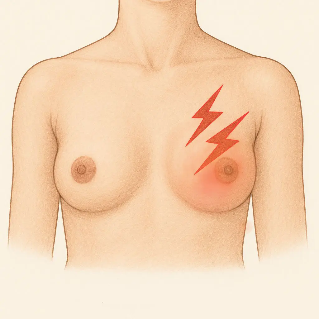
Breast pain
Usually, this is a diffuse pain related to the cyclic changes that occur during the menstrual cycle. It usually involves both breasts and can be associated with swelling or nodularity. It typically occurs 1-2 weeks before the onset of the period and subsides with menstrual bleeding. Interventions are generally not required; however, pharmacological therapies can be applied according to the severity of the condition.
Non-cyclic breast pain tends to be limited to a localized area. These are less related to hormonal causes but rather due to certain anatomical factors. The causes may involve trauma, ruptured cyst, inadequate breast support, inflammation of the breast, non-cancerous tumours, mammary duct ectasia, pregnancy or the effect of some medications. Breast pain can also be due to extramammary causes like cardiac, musculoskeletal or gastrointestinal pathologies.
Women presenting with breast pain without obvious cause are referred for breast imaging( ultrasound or mammogram according to her age) for identification of the cause. Almost all cases present with pain are benign, especially in the absence of palpable masses or any other abnormal findings. However, about 10% of breast cancers can cause pain.
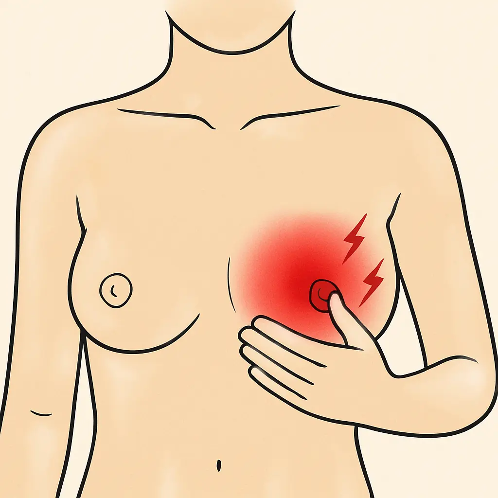
Redness, swelling, heat and pain
These are the characteristic features of breast inflammation. (mastitis) It is a relatively rare condition that occurs mostly during the first weeks of breastfeeding. (lactational mastitis) Pathogenic bacteria( staphylococcus aureus) gain entry via the nipple,
* form pus-filled cavities(abscess)
* may cause pus discharge following rupture
* nipple discharge
It can associated with
* painful nodules at axilla( axillary lymphadenopathy),
* fever.
This is effectively treated with antibiotics, and in some cases, drainage may be needed. The following information is essential regarding the continuation of breastfeeding.
* Feeding from the affected breast is not harmful to the baby. Although some bacteria can pass through breast milk, mother is passing enough amount of antibodies that protect the baby.
* Should ensure that milk does not contain pus or blood.
* Expression of milk from the affected side is essential.
* Mother can feed the baby from other breast following drainage, but milk should be manually removed.

But if a non-lactating, older woman presents with breast inflammation, it may be alarming. Inflammatory carcinoma of the breast, present with
* Swollen, reddish breast
* No identifiable underlying lump
As this type of cancer can spread easily, it is important to seek medical care early.
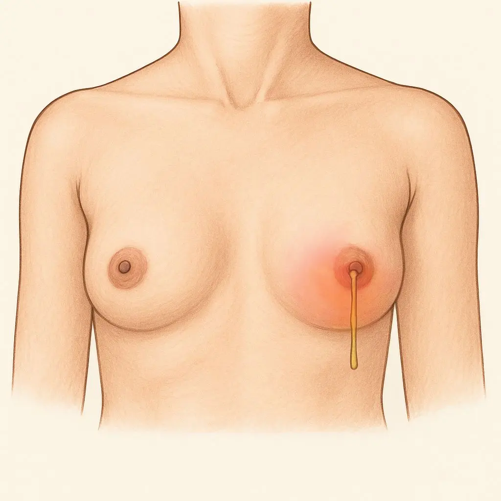
Nipple discharge
Nipple discharge refers to the leaking of fluid from one or both breasts in non-pregnant or non-breastfeeding women. This is the third leading complaint in women present with breast pathologies. Often, this may be a benign condition(>97%).

Physiological causes for nipple discharge include,
* Spontaneous or intentional termination of pregnancy
* Hormonal fluctuations associated with menstrual cycle
* Discharging of breast milk(galactorrhoea) may last up to two years following delivery
* Intraductal papilloma
This is the most common benign cause of nipple discharge. Produce unilateral, clear, serous discharge or bloody discharge from large ducts below the nipple. Usually, discharge comes out of a single duct. Removal of the affected duct improves the condition.
* Duct ectasia
This is a benign condition that causes dilated and tortured lactiferous ducts. It is associated with periductal inflammation and fibrosis. Patients present with nipple discharge in varying colours (white to grey/black to grey). It can be associated with pain around the nipple and areola, inversion of the nipple, and a palpable lump below the areola. It mimics breast carcinoma due to this clinical picture. If you are experiencing these symptoms, it is essential to seek medical attention to determine the underlying cause.
If it's confirmed as duct ectasia,
[  ] Just be relaxed. It's nothing dangerous
[  ] Apply warm compresses over the middle part of the breast
[  ] Wear a supportive bra with pads that can absorb discharges
[  ] Maintain general hygiene
[  ] Take antibiotics as described if there is overlapping inflammation.
* Fibrocystic changes in the breast
These are benign lesions, including cysts, fibrosis, adenosis, etc. Occur due to high estrogen state in women at the age of (25-45).

Physiological or normal conditions mostly lead to clear, serous discharge, while pathological conditions lead to the discharging of bloody, purulent discharges. These conditions include
* Breast infection and abscess
* Malignant conditions of the breast
Some cancer types, including intraductal carcinoma and Paget disease of the breast, can cause nipple discharges. Paget disease is a manifestation of ductal carcinoma in situ, the earliest stage of ductal carcinoma. It presents with crusted exudate over the nipple and areola.
So, all women present with nipple discharge, mainly older women after cessation of menstruation, are investigated for possible underlying malignancy.
* Pituitary tumour causing prolactinoma
This leads to abnormal milk production in non-pregnant women.
* Systemic diseases that elevate prolactin levels ( disorders in the pituitary or hypothalamus,  chronic kidney disease, renal disease )
* Side effects of some medications (oral contraceptives, opioids, antihypertensives)
Palpable masses
More than 95% of palpable masses are benign. You can palpate the noticed mass to evaluate its size, shape, margins, consistency, surface and attachment.
Benign tumours tend to be round or oval in shape, having regular margins and a smooth surface over the lump. Fibroadenoma, which is commonly found in young females, is a better example of this. They are usually single or multiple nodules that are not hard (firm) and have no attachments with skin or underlying muscles. However, there are some cancers, such as mucinous carcinoma, that can mimic benign tumours. So, you can't confirm whether it is benign or malignant only by palpation.

On the other hand, malignant masses usually exhibit the following features.
* Painless mass
* Size increases with time
* Nipple discharge, mostly bloody
* Inward pulling of skin (skin tethering)
* Dimpling over skin (like an orange peel)
* Mass may be less mobile (attached to skin or underlying muscles)
* May present with painless nodules under the armpit (axillary lymphadenopathy)
* Having irregular borders
* Surface over mass is rough
If you are presenting with this type of mass, seek medical advice immediately.
There are many identified risk factors for breast cancer. Those are,
* Old age and female gender
More than 75% of women with breast cancer are older than 50 years. Males are getting only 1% risk compared to females.
* Family history of breast cancer
* Diet, reproductive patterns and breastfeeding practices of a population
* Long exposure to estrogen
This includes getting their first menstrual period early (early menarche), late menopause, women not having given birth(nulliparity), absence of breastfeeding and older age of first pregnancy.
* Exposure to ionizing radiation
* Other risk factors like post-menopausal obesity, hormone replacement therapy and alcohol consumption

 If you have these risk factors, you may be more vulnerable to some malignant condition of the breast. A monthly self-breast examination is recommended for all women above 20 years of age, one week after initiation of menstrual bleeding, to identify any abnormality of the breast. Screening of females is also done at the community level. Early detection of breast cancer gives better outcomes. This guide provides you with only basic knowledge. If you detect some abnormal finding, never delay to seek medical care.